


Bone Mineral Density
Bone density measurement or bone densitometry is the name of a method that can be used to determine the stiffness and degree of strength of the bones in the body. There are different ways to measure bone density, but the most common and easiest method is to use the DEXA method. Bone density is measured at the Taba Imaging Center with the most up-to-date equipment. Measuring bone density is completely painless and does not require any prior preparation.
Bone mineral density is measured daily at the Taba Center from 8 am to 10 pm without the need for an appointment.
Bone mineral density is measured daily at the Taba Center from 8 am to 10 pm without the need for an appointment.
MRI
MRI 1.5 tesla GE USA
Large tunnel
In most imaging, the patient's head is placed outside the tunnel.
Overweight patient imaging is possible
Silent MRI
Removal of metal and motion artifacts
3D MRI imaging
Diffusion and perfusion MRI
MR Angiography without injection
Large tunnel
In most imaging, the patient's head is placed outside the tunnel.
Overweight patient imaging is possible
Silent MRI
Removal of metal and motion artifacts
3D MRI imaging
Diffusion and perfusion MRI
MR Angiography without injection
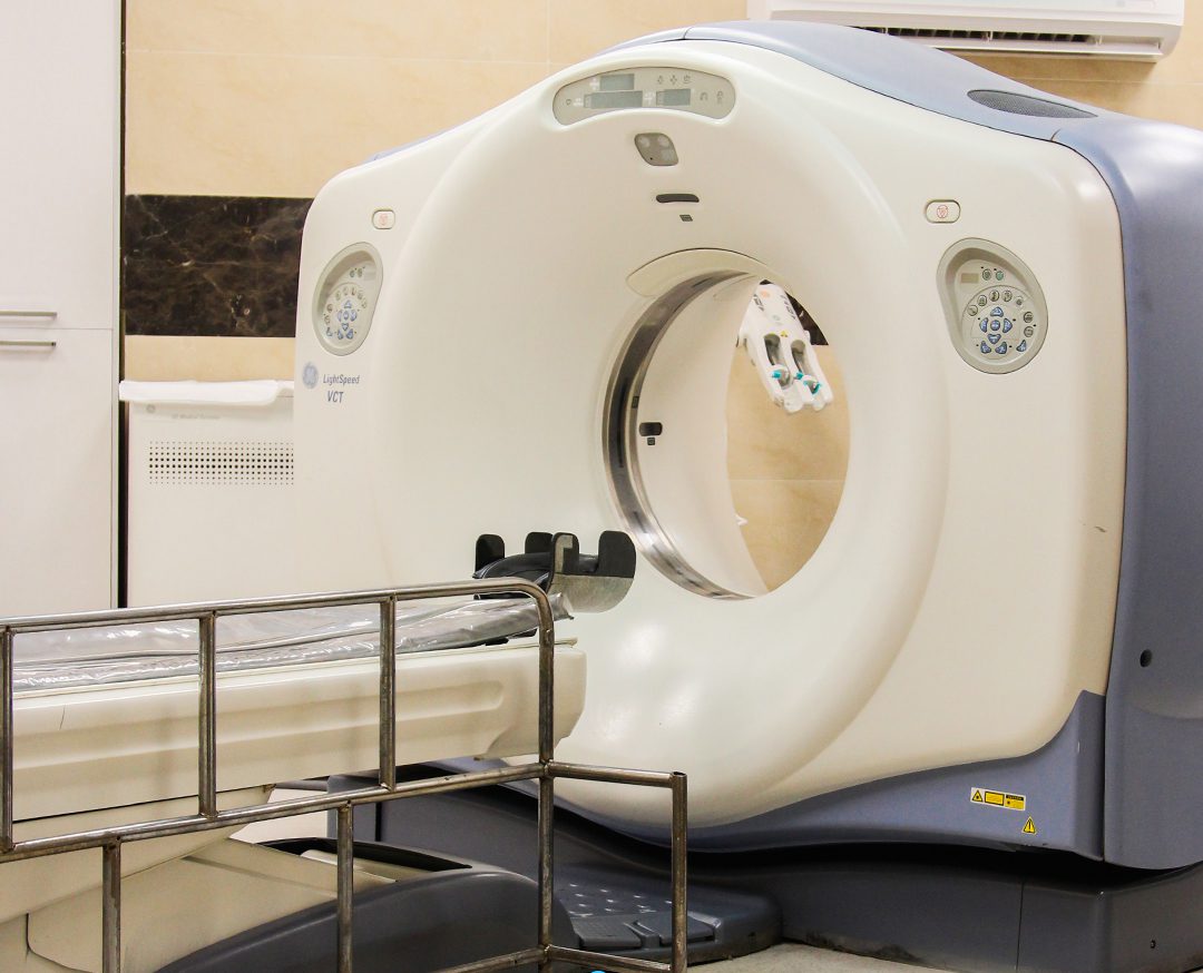
CT Scan
CT scan is performed using x-ray radiation, but in this center, using the advanced CT scan machine of 128 slices GE USA, the level of radiation received by the patient’s body is reduced by 70%.
CT scan images are more accurate in showing the shape of bones and rough structures of the body compared to radiography images.
Using this technique, the inside of the bone can also be seen.
CT scan images are more accurate in showing the shape of bones and rough structures of the body compared to radiography images.
Using this technique, the inside of the bone can also be seen.
A variety of CT Angiography methods, including CT coronary angiography, are performed daily at the Taba Medical Imaging Center.

Mammography
Mammography is performed by a digital medical device with minimal pain and reduced radiation.
Mammography is performed by a digital medical device with the least pain and reduced levels of radiation. According to research, breast cancer is one of the most common cancers among women. It is recommended that women over the age of 40 have an annual mammography test to diagnose breast tumors. Early detection of breast cancer with mammography and effective treatment strategies play an important role in the healing and elimination of breast tumors.
Mammography is performed by a digital medical device with the least pain and reduced levels of radiation. According to research, breast cancer is one of the most common cancers among women. It is recommended that women over the age of 40 have an annual mammography test to diagnose breast tumors. Early detection of breast cancer with mammography and effective treatment strategies play an important role in the healing and elimination of breast tumors.
Sonography
Ultrasound is safe and painless and produces images of the inside of the body using sound waves. This imaging involves the use of a small transducer (probe) and gel that is placed directly on the skin.
This medical imaging device involves the use of a small transducer (probe) that is placed directly on the skin. Ultrasound examinations do not use harmful radiation (as used in X-rays), so it is not dangerous for the patient.
This medical imaging device involves the use of a small transducer (probe) that is placed directly on the skin. Ultrasound examinations do not use harmful radiation (as used in X-rays), so it is not dangerous for the patient.
Ultrasound is performed daily at the Taba Center with 3 up-to-date devices.
Digital Radiology
At the Taba medical center, imaging is performed using modern devices, which has a significant impact on the final image quality. The use of devices that can move in all direction allows a seamless process for patients with limited movement ability.
### The importance of using digital devices is especially evident in lumbar vertebrae. ###
### The importance of using digital devices is especially evident in lumbar vertebrae. ###